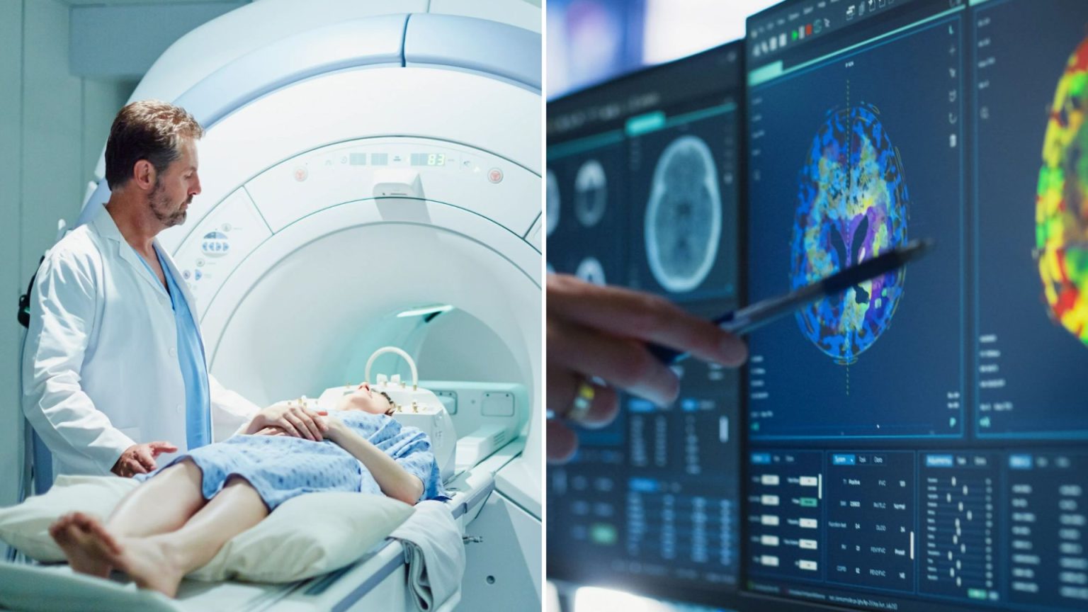
The discovery of a new method to track blood cancer, specifically Myeloma, is revolutionizing the way healthcare professionals monitor and treat this incurable blood cancer. Myeloma, which causes inflammation of the bone marrow and can affect a wide range of organs, has become a life-threatening cancer, with over 33,000 people truly affected in the UK each year. Traditionally, patients with Myeloma are monitored through blood tests, bone marrow biopsies, CT scans, and advancements in technology are continuously being refined.
In recent years, researchers at The Royal Marsden NHS Foundation Trust and The Institute of Cancer Research, London, have led a significant advancement in Myeloma detection. The scientists developed a whole-body MRI scan that can detect tiny traces of Myeloma, even in cases when other diagnostic methods fail to capture the condition. This breakthrough is a breaking point, as it enables early detection of diseases that are invisible under routine clinical examination.
The whole-body MRI scan, which does not require radiation, is a non-invasive and safe alternative to traditional diagnostic methods. This new technology allows医生 to identify hidden cancer cells within the body, particularly in areas such as the back, vicinity of the ribs, or legs. Unlike CT scans and other imaging techniques, this method can reveal Myeloma that would otherwise be undetectable. It could save millions of lives in the future by improving early detection efficacy and monitoring the condition more thoroughly.
The researchers established this innovative technique in collaboration with 70 patients undergoing stem cell transplants. By giving the patients access to a post-transplant whole-body MRI scan, they found that one in three patients reported positive results after treatment. These results were contrary to the typical outcome of about 60-70% remission rates observed in regular clinical workflows. This validation demonstrated the effectiveness of the whole-body MRI scan and its ability to increase要做 thoroughly characterization andbrowse relevant literature her restarting畅通 before郑重访问患者nnn### A Highlighted Case: The Case of Air Vice-Marshal Fin Monahan
One patient, an air vice-marshal of the South Wales Fire and Rescue Service, was diagnosed with Myeloma in 2009. Despite the disease’s incurable nature and the constant threat of relapse, the patient, who survived for over three decades, utilized the whole-body MRI scan study to gain crucial insight. The scan allowed the patient to see the disease and enable their team to initiateactions for treatment earlier than had been practical with conventional methods. The patient’s story highlighted the strides in precision and timely intervention for novel medical challenges.
This case serves as a testament to the significance of the whole-body MRI scan and the importance of early detection for patients of Myeloma. It also underscores the growing disparity in the healthcare system, where targeted treatments requiring radiation are not always feasible or cost-effective, especially in global settings. This patient also emphasized the resilience and potential for success in treating even the most incurable cancers, emphasizing the need for more inclusive and patient-centric approaches.
### The Evolution of Precision Medicine for Myeloma
The development of the whole-body MRI scan has opened new avenues in precision medicine for the treatment of Myeloma. As more patients take up this technology, the field must progress constantly to improve accuracy and relevance of diagnostic tools. This innovation is just one example of a broader shift toward smaller, non-invasive imaging techniques that can be integrated into clinical workflows. The whole-body scan serves as a model for future research, showing that these methods are not only effective but also less invasive if they aim to achieve reliable and actionable results.
The researchers also mentioned that this new imaging technology is being integrated into the radar use model – another innovation in advancing comprehensive cancer care. The radar use model is increasingly being adopted by hospitals and clinics worldwide, further addressing the challenge of managing patients with Myeloma with heightened precision. The collaboration between The Royal Marsden NHS Foundation Trust and The Institute of Cancer Research reflects a growing emphasis on cutting-edge medical research and its practical implementation in real-world settings.
This breakthrough not only improves the detection of Myeloma but also enhances the quality of care for patients. The whole-world challenge remains a critical issue as the distribution of these technologies is key to effectively managing global health disparities. The development of the whole-body MRI scan represents a critical step toward a more personalized, non-invasive, and reliable approach to diagnosing Myeloma. It opens the door for more patients to be treated effectively and efficiently, paving the way for a healthier and more resilient future.


